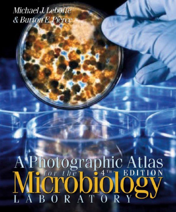
Imagine peering into a breathtaking landscape, a world teeming with life, yet invisible to the naked eye. This microscopic panorama is the domain of histology, the study of tissues and their intricate structures. It’s a realm where colours burst forth, shapes dance in vibrant patterns, and the building blocks of life unfold in astonishing detail. To explore this hidden world, we turn to a crucial tool: a photographic atlas of histology PDF.

Image: www.carousell.ph
A photographic atlas of histology PDF acts as a virtual window into the microscopic world, showcasing the diverse array of tissues that make up our bodies. It’s a treasure trove of detailed images, descriptions, and explanations, bringing clarity to the complex tapestry of human biology. Whether you’re a student embarking on a journey of medical discovery or a seasoned professional seeking a comprehensive reference, a photographic atlas of histology PDF is an invaluable companion.

Image: medicalstudyzone.com
A Photographic Atlas Of Histology Pdf
Delving into the Microscopic Tapestry: A Photographic Atlas of Histology PDF Unfolds
The journey into histology starts with the fundamental building blocks of life: cells. These tiny units, each a marvel of nature, organize themselves into tissues, forming the foundation for all organs and systems. A photographic atlas of histology PDF captures these intricate arrangements, revealing the diverse tapestry of tissues that make up our bodies.
Epithelial Tissue: The Guardians of Our Surfaces
Imagine a protective shield, a barrier separating the inside from the outside, constantly working to maintain balance. This is the role of epithelial tissue, lining the surfaces of our bodies, from the skin to the lining of our internal organs. A photographic atlas of histology PDF reveals the fascinating variety within epithelial tissue, showcasing the tightly packed cells arranged in sheets, some thin and delicate, others thick and resilient, each playing a crucial role in shielding and regulating our internal environment.
Connective Tissue: The Scaffolding of Life
Imagine a network of support, a framework that binds and provides structure, allowing movement and resilience. This is the realm of connective tissue, the most diverse of tissue types, providing support, protection, and structure throughout the body. A photographic atlas of histology PDF dives into this intricate world, revealing the various forms of connective tissue, from the dense and fibrous tendons to the delicate and pliable cartilage, each playing a vital role in maintaining the body’s form and function.
Muscle Tissue: The Movers and Shakers
Imagine the grace of a dancer, the strength of an athlete, the beating of a heart – these are all examples of the work of muscle tissue. A photographic atlas of histology PDF allows us to explore the fascinating world of muscle tissue, revealing the microscopic structures that enable movement, from the smooth contractions of the digestive tract to the voluntary movements of our limbs.
Nervous Tissue: The Communication Network
Imagine a vast network, a web of interconnected threads, transmitting information at lightning speed. This is the essence of nervous tissue, the master communicator of our bodies. A photographic atlas of histology PDF guides us through this incredible world, showcasing the intricate structures of neurons, the highly specialized cells that transmit signals throughout the body.
Embarking on a Visual Journey: The Advantages of a Photographic Atlas of Histology PDF
A photographic atlas of histology PDF offers a unique advantage: the power of visual learning. Our minds are wired to retain information more effectively when it’s presented visually, and a photographic atlas leverages this natural tendency. Each image becomes a story, a window into the microscopic world, allowing us to grasp concepts and nuances that written descriptions alone can’t fully convey.
Beyond the Images: The Value of Descriptions and Annotations
A photographic atlas of histology PDF is more than just a collection of images. It’s a comprehensive resource, providing detailed descriptions and annotations to complement the visuals. These written explanations offer context, clarify key features, and delve into the functions and complexities of each tissue type.
Unlocking the Power of Knowledge: Using a Photographic Atlas of Histology PDF for Success
For students, a photographic atlas of histology PDF is an invaluable tool for grasping complex concepts and visualizing microscopic structures. It serves as a visual companion, enhancing understanding and retention. For professionals, it’s a readily accessible reference, providing a comprehensive overview of tissues and their characteristics for diagnosis, research, and clinical practice.
Embracing the Microscopic World: A Journey of Discovery
A photographic atlas of histology PDF is a gateway to the wondrous world of microscopic structures. It’s a resource that unlocks understanding, empowers learning, and inspires curiosity. Whether you’re a student, a professional, or simply someone fascinated by the intricacies of life, a photographic atlas of histology PDF offers a captivating journey into the fascinating world of tissues and their role in the grand symphony of life.


